


 Play store

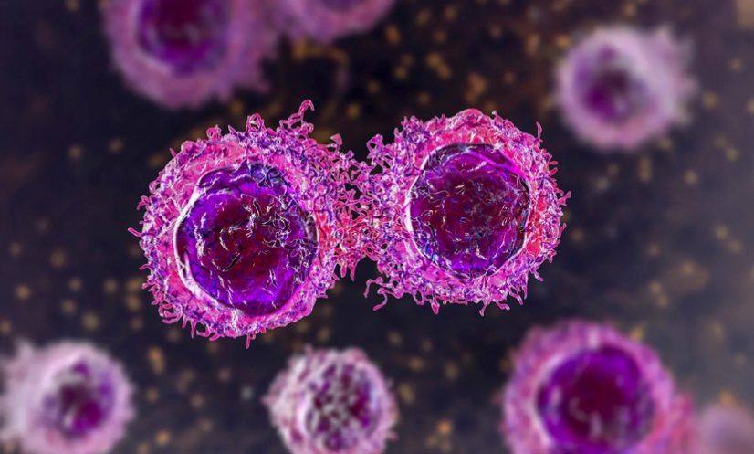
Researchers have identified environmental conditions that may help cancer cells build a sugar-rich protective coating that makes them harder for the immune system to detect and destroy.
The study, led by researchers at the Sanford Burnham Prebys Medical Discovery Institute and collaborating institutions across North America, was published in Science Advances on August 7, 2026.
The findings suggest that changes in the tumour microenvironment—the surrounding network of immune cells, connective tissue, blood vessels, proteins and carbohydrates—can influence how effectively cancer cells conceal themselves.
The researchers also identified a potential way to reduce the coating, raising the possibility of making cancer cells more vulnerable to immune attack.
Tumour environment alters cancer cells
Lead and corresponding author Kevin Tharp, PhD, said he became interested in the role of physical forces after studying how pressure on cells can alter mitochondrial function.
“Primary tumours are typically stiffer than their surrounding tissue,” said Tharp, an assistant professor in the Cancer Metabolism and Microenvironment Program at the Sanford Burnham Prebys NCI-Designated Cancer Center.
“This led me to hypothesize that the biophysical properties of cells influence the altered metabolic programs that everyone observes in tumours.”
To test the idea, the researchers exposed cells to abundant glucose under different physical and nutritional conditions.
Some cells were grown in stiff environments designed to mimic primary tumours, while others were placed in softer environments resembling normal tissue. The cells were also grown in either conventional laboratory media or a formulation designed to more closely reflect the nutrient composition of the human body.
Both groups were tested under normal and elevated glucose conditions to simulate hyperglycaemia.
High glucose thickens sugar coating
The different conditions produced significant changes in the proteins and metabolites inside the cells, as well as in the thickness of a sugar-derived layer covering their surfaces, known as the glycocalyx.
Notably, excess glucose increased the thickness of the glycocalyx only when cells were grown in the physiological medium designed to mimic conditions inside the human body.
The researchers found major differences in the glycoconjugates—carbohydrates attached to proteins or lipids—produced by cells grown in conventional media compared with those grown in physiological media.
They also found that hyperglycaemia altered the composition of these glycoconjugates.
Because glucose provides some of the raw material required to produce glycoconjugates, the researchers suspected that changes in glucose metabolism could influence the formation of the glycocalyx.
“We observed that changing the physiological media composition and changing the available metabolites for those tumour cells reveals distinct biology for normal and tumour cell metabolism,” Tharp said.
HSF1 emerges as key player
The team then examined proteins that became more abundant when cells were exposed to high glucose. Their experiments highlighted heat shock factor 1 (HSF1), a protein that helps cells respond to heat and other forms of stress.
Previous studies have also linked HSF1 to breast cancer progression and metastasis.
The researchers found that the presence or absence of HSF1 altered the composition of glycoconjugates produced by the cells.
They subsequently investigated how hyperglycaemia, HSF1 and the tumour microenvironment interact to affect the immune system’s ability to recognise and attack cancer cells.
The findings point to the tumour environment and cancer-cell metabolism as potentially important targets for weakening the protective glycocalyx and improving immune recognition of cancer cells.